
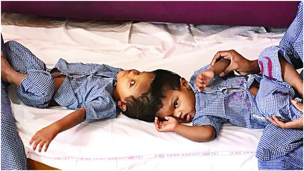
Two 8-hour long rehearsals on polylactic acid model saved craniopagus twins at AIIMS
This was the first time in the country that a 3D model was used for practice. The polylactic acid model was based on the information from the MRI, CT-Scan, and Angiogram of the twins and resembled their head structures. "This model helped us as we knew precisely which parts had to be operated upon and where to put incisions. The model had Jagga and Balia marked on the heads for us to know," said Dr Deepak Gupta, Pediatric Neurosurgeon, AIIMS. While several head graftings (transplants) have been performed on them, the twins still miss skull bones and have to undergo the last round of surgery.
Source: dna February 12, 2018 00:33 UTC